CT Scan vs MRI Brain Tumor: 7 Key Facts for Emergency Care
Introduction
Medical imaging is super important for doctors trying to spot brain tumors. When it comes to imaging tech, especially in hospital emergency rooms, CT scans and MRI brain tumor imaging are the big two. These tools help find, pinpoint, and see how bad any weird stuff is in the brain. Knowing the differences between them helps patients, caregivers, and even med students figure out which test is best in different cases. Both CT and MRI scans have good and bad sides, which come into play in emergency medical care. Here are seven facts showing how different imaging methods give fast, spot-on diagnoses, which affects how patients are treated and how well they recover.
1. The Fundamental Difference Between CT and MRI Scans
Both CT scans and MRIs let doctors see inside your body, but they do it in different ways. CT scans use X-rays to quickly snap pictures of your brain – They’re good for seeing quick details. MRIs take more time but give way more info about brain tissue, what tumors are, and what’s around them. This helps doctors decide what to do in a brain emergency.
2. Speed and Accessibility in Emergency Situations
Time is critical when treating patients in an emergency room. These patients can come in with different acute neurologic symptoms such as severe headache, changes in mental status, seizure activity, or even loss of consciousness. Computed tomography has some benefits over magnetic resonance imaging in this case: it is almost always available in emergency departments; it can be done quickly; and the results usually come back within minutes. Furthermore, MRI machines are not as common as CT machines found in emergency departments. For quick evaluation of possibly life-threatening conditions such as intracranial bleeding or large mass effects, CT is usually the first choice for imaging because it works fast and can check these important problems immediately.
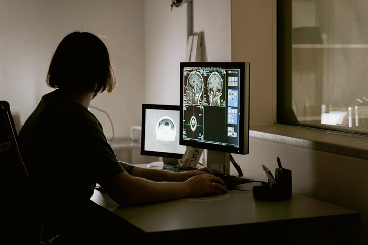
3. Accuracy and Detail in Tumor Identification
MRI is the gold standard for detecting benign and malignant brain tumors due to its ability to visualize soft tissues with high resolution radiologists can detect even the smallest changes. It also gives information on the tumor’s volume, location, and proximity to neighboring structures which is very helpful in precise neurosurgical planning. Despite the fact that CT scans are excellent for evaluation of dense tissues and hemorrhages, they might not be as successful in detecting very small changes in soft tissue. However, with the use of contrast agents, CT can still reveal the tumor’s blood supply and calcifications and thus retain its value as a diagnostic tool. Actually, both practices are common: CT is utilized for rapid initial evaluation in urgent cases while MRI is reserved for extensive diagnosis and follow-up.
4. Radiation Exposure and Patient Safety
CT and MRI have a lot of differences among which one of the most important is regarding the issue of patient safety in terms of radiation. CT scan uses ionizing radiation, and its dangerous effects can be seen in children and pregnant women if continuous exposure occurs. On the other hand, MRI is not using any radiation at all which thus is a less risky method for monitoring over a long period of time or conducting multiple scans.
In emergency situations, however, the advantage of a fast CT scan is so much greater than the slight radiation risk, particularly when the patient’s life is in danger. Doctors take each case very carefully and weigh the diagnosis’s urgency against safety issues. The safety benefit of MRI is most pronounced in follow-up imaging where detailed monitoring of tumor development is being done without the concern of cumulative radiation exposure.
5. Cost and Availability Across Medical Facilities
Another major factor in decision-making is the cost and availability. CT scanners require much less cost of operation and are found in hospitals more often, especially in the case of smaller cities or underdeveloping areas. Their fast processing time and lesser cost of operation put them forward as the best option for initial evaluations in emergencies. MRI machines on the other hand, because of their complicated structure and high maintenance, are more expensive and not so common. This situation can interfere with the access to first-class imaging in some places. Yet, the MRI, when present, is still the best option in terms of precision in detecting brain tumors. The use of both technologies together provides full care – CT for emergency triage and MRI for detailed diagnostic confirmation.
6. Role of CT and MRI in Emergency Care
In Emergency Care, CT and MRI are both important tools but serve different purposes. In fact, CT scans are generally the initial exams that are performed when a patient arrives at the emergency department with acute neurological symptoms. They fast detect bleeding, fractures, or edema, which might require urgent surgical intervention, a.k.a. cutting the patient open. Subsequently, MRI is performed once the condition of the patient is stabilized to deliver additional details such as ascertaining whether it is a tumor, infection, or a demyelinating disease that has caused the patient’s symptoms.
MRI’s precision allows clinicians to tell the different tumor types apart, to evaluate the degree of damage to the brain tissue, and to organize the treatment accordingly. On the other hand, the combined use of CT and MRI ensures that the emergency teams are able to respond effectively in both short and long terms hence, maximizing the patient’s outcome and improving the chances of survival in critical scenarios.
7. The Use of Contrast Agents in Brain Imaging
Both methods may utilize contrast materials to make the images clearer and to differentiate between healthy and unhealthy tissues. In the case of CT scans, iodinated contrasts bring blood vessels and tumors into focus, while gadolinium-based contrasts in MRI show the internal structure of brain lesions more clearly than ever before. Nevertheless, the use of contrast agents is restricted to certain conditions only, primarily for patients with kidney problems or those having allergies. Furthermore, MRI contrast gives a better understanding of the tumor anatomy and its connection to brain operation, hence it is the favored technique for follow-up studies. In acute situations where quick results are required, CT contrast is still beneficial for fast and precise finding of abnormal tissue growths or determining hemorrhage patterns.
8. Detecting Tumor Complications and Post-Treatment Monitoring
CT scans and MRIs are both critical in spotting the tumor’s complications such as swelling, bleeding, or even death of tissue after the treatment. One of the main benefits of radiology is that it makes it easier to visualize the recovery of tissues and so evaluate the success of radiation or chemotherapy. The fastness of CT scans makes it possible to use them in emergencies where rebleeding or an increase in intracranial pressure is suspected.
In the case of post-surgical situations, the use of MRI helps in the identification of tumor cells or the case of recurrence. It is the combination of all these modalities that allows for the complete tumor management. All of them are used in the continuum of care—from emergency detection to long-term follow-up—ensuring accuracy and patient safety throughout.
9. Choosing Between CT and MRI: Clinical Decision Factors
The selection of imaging modality, CT or MRI, is influenced by a lot of factors like the patient’s condition, urgency of the matter, and the clinical goals. In the case of trauma or suspected bleeding, CT is the go-to choice due to its quickness and ability to depict the dense structures clearly. However, MRI is still the best choice for soft-tissue evaluation, tumor study, or surgical planning.
Doctors still rely on the patient’s symptoms, the availability of resources, and the risk involved to make their choice. Take for instance; a patient with a suspected brain hemorrhage will be the first one to get a CT, whereas, just the opposite, the one with neurological symptoms for quite some time could be counseled to undergo an MRI for more detailed insights. Such a balanced approach ensures that the selected imaging technique not only meets the patient’s immediate needs but also serves the long-term diagnostic requirements.
10. The Future of Brain Imaging in Emergency Medicine
The modernization of technology is very slowly but surely changing the whole scenario of brain imaging. The systems that combine different technologies like PET-MRI and AI-powered diagnostic tools are now giving a big boost to the accuracy of images and the speed of interpreting them. It is possible that in the not-too-distant future, hospitals or emergency services will have AI-powered systems that can automatically decide if the patient should go for a CT or MRI based on the doctor’s analysis and symptoms.
In addition, there is a growing interest in portable MRI which is expected to provide access even in the case of the smallest hospitals and rural clinics. The progress of emergency medicine will still rely on the combination of the two techniques—quick CT and detailed MRI—to set the bar for brain tumor evaluation. The future looks bright with quicker, safer, and more precise diagnosis that do not compromise on either speed or accuracy.
Conclusion
The ongoing debate around CT Scan vs MRI Brain Tumor has not, so far, changed the importance of both imaging methods, especially in emergencies. CT scans are very fast and are the best option when an immediate diagnosis is needed. MRI, on the other hand, gives the necessary detailed clarity for complete tumor analysis. Their combined use guarantees the best quality care—from emergency response to long-term treatment planning. The continuous evolution of technology will be by the integration of both techniques, thus, getting more accurate diagnoses, enhancing survival rates, and reinforcing the foundation of modern medical imaging.
In the end, the aim is no longer to select the one between CT and MRI but to realize how the two can work together in diagnosing, treating, and monitoring brain tumors under the broader aspect of Emergency Care.